
Otosclerosis is a progressive condition affecting the bony labyrinth of the inner ear, leading to hearing loss. It is characterized by abnormal bone remodeling, primarily around the stapes footplate, which can result in stapes fixation and impaired sound transmission. Audiological evaluation plays a crucial role in diagnosing and understanding the extent of hearing loss in otosclerosis.

Key Audiological Findings
Conductive Hearing Loss:
The hallmark of otosclerosis is conductive hearing loss, particularly in its early stages. This occurs due to the fixation of the stapes, which hinders the transmission of sound from the middle ear to the inner ear.
The hearing loss typically begins in the low frequencies and progresses to involve higher frequencies as the condition advances.

Carhart's Notch:
A distinctive feature in the audiogram of otosclerosis patients is Carhart's notch, observed as a dip in bone conduction thresholds around 2000 Hz. This is attributed to the mechanical effects of stapes fixation on cochlear function.

Air-Bone Gap:
Pure-tone audiometry often reveals a significant air-bone gap, indicative of conductive hearing loss. In advanced cases, the gap may widen, reflecting the severity of stapes fixation.
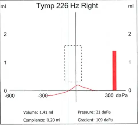
Impedance Audiometry:
Tympanometry typically shows normal middle ear pressure but reduced compliance, reflecting the stiffness of the ossicular chain.
Acoustic reflexes are often absent or elevated due to the fixation of the stapes.
Mixed Hearing Loss:
In cases of cochlear otosclerosis, where the condition affects the cochlea, a sensorineural component may be added to the hearing loss. This results in a mixed hearing loss pattern on the audiogram.
Speech Audiometry:
Despite significant hearing loss, speech discrimination scores are often excellent in patients with otosclerosis, unless there is cochlear involvement.
Diagnostic and Clinical Implications
Audiological findings not only aid in diagnosing otosclerosis but also guide treatment decisions. For instance:
Hearing Aids: Patients with conductive hearing loss may benefit significantly from amplification devices.

Surgical Intervention: Procedures like stapedotomy or stapedectomy can restore hearing by bypassing the fixed stapes.

Monitoring Progression: Regular audiological evaluations help track the progression of the disease and the effectiveness of interventions.
In conclusion, audiological assessments provide critical insights into the nature and extent of hearing loss in otosclerosis. Early diagnosis and appropriate management can significantly improve the quality of life for affected individuals.
Comentários